Precision-cut tissue slices are thinly and identically cut sections of living people`s tissues that contain native cellular diversity, natural extracellular matrix, and preserve the physiology of the real organ.
These tissue slices are used in a wide range of biomedical studies, including drug/chemical metabolism and penetration measurements, the development of gene therapy technologies, infection and cancer modelling, and personalized medicine research.
Therefore, it is important to determine biopsies` best microenvironment to keep them active and alive.
A biopsy is a collection of millions of cells, and when incubated simply in a complete medium in a well, it does not replicate the tissue microenvironment. The biopsy is exposed to an uneven nutrient gradient and suffers loss of structure as it lacks external support.
Human ECM sol-gel system (HECM) imitates human ECM, which is made up of proteins such as collagen, elastin, laminin, and fibronectin. Tissue slices integrate well within HECM hydrogel, maintaining higher levels of viability than supported by the typical gel-like materials, such as Matrigel or collagen-based hydrogels. HECM creates a network of proteins associated with the biopsy that support its organization, creating a semi-permeable barrier in which all cells are in the same conditions. The gel also forms a protein mesh into which cells can easily migrate from the tissue.
We offer a simple and effective protocol for creating models from biopsy and HECM.
Protocol steps:
1. Dilute HECM in the specified ratio in ice-cold medium. Mix carefully, use a vortex or a cooled ultrasonic bath for better results. We recommend the use of 1/20 dilutions for better results.
2. Pour the required amount of the prepared solution slowly into the wells. For dilution 1/20, we recommend 100 µl of diluted HECM /well for a 96-well plate.
3. Carefully tilt the plate from side to side to let the liquid cover the entire surface of the well.
4. Centrifugate the plate at 4°C, 2500 g, 10 minutes.
5. Incubate the plate at 37°C overnight, avoiding any agitation to prevent disturbing the gel.
6. While the incubation continues, prepare the tissue biopsy.
7. After incubation, aspirate any excess liquid from the wells.
NOTE: Avoid touching the bottom of the well with tips, as it will damage the coating. Do not allow the HECM to dry.
8. Place a tissue piece on the coating. Leave in the incubator for 10 minutes
9. Add 50 µl of HECM in a dilution ratio of 1/5-1/10 on top of the inserted biopsy pieces and incubate the assembled tissue culture for a minimum of 1 hour.
10. Carefully add 150 µl of warm culture medium on top of each culture well scaffold and incubate overnight.
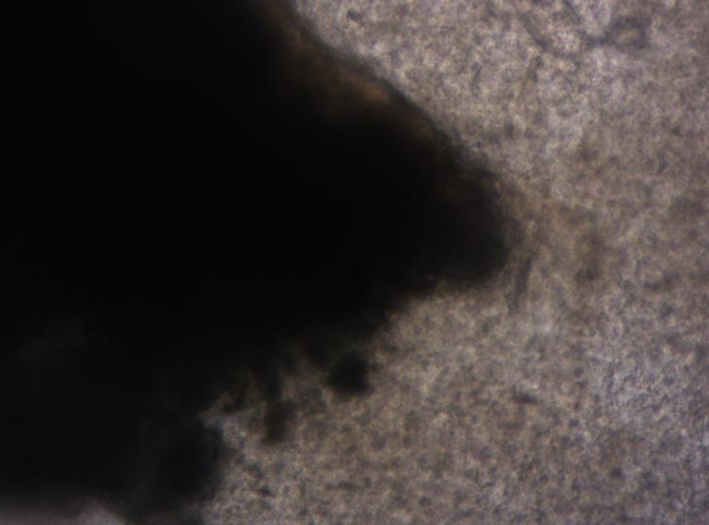
We proved that the viability of precision-cut tissues slices incubated for three days surrounded by gel is much better than in medium alone by staining cells with Alamar Blue.
| Dilution of HECM | Results of spectrum reading OD |
| 1/5 | 11166 |
| 1/10 | 18316 |
| 1/20 | 15817 |
| No HECM | 13698 |
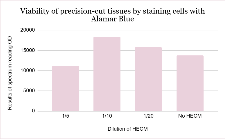
AlamarBlue functions as a cell health indicator, enabling quantitative measurement of cell viability and proliferation using absorbance- or fluorescence-based microplate readers. It uses the natural reducing power of living cells to convert resazurin to fluorescent resorufin.
HECM hydrogel mimics physiological conditions for a biopsy, so when used at a 1/5 dilution, the matrix is denser than the native one. It may limit oxygen diffusion, nutrient transport, and waste removal.
Higher values of the spectrum reading OD of biopsies coated in 1/10 and 1/20 dilutions of HECM gel show that they have more alive cells, which matches the expected results.
Explore our product catalog to see how HECM can support your specific tissue types.