


How well do current models predict human drug response?
Human ECM in drug development is becoming a critical tool for closing the gap between traditional cell culture and real human biology.
Traditional drug discovery is a long and expensive process that can take 10–15 years. After target validation, compounds usually pass through high-throughput screening, lead optimization, animal testing and finally clinical trials. The compound may look promising in early in vitro testing, perform differently in animals and then fail once it reaches human trials.
One of the most widely used systems is conventional 2D cell culture. It is simple, fast, inexpensive and compatible with high-throughput screening. However, cells on plastic grow on a rigid surface that does not reflect the architecture, stiffness or matrix signals present in real tissues.
Three-dimensional models were developed to close part of this gap.
Spheroids are cell aggregates that improve cell-cell interactions and generate gradients of oxygen. However, spheroids do not reproduce tissue-specific organization or cellular diversity.
Organoids offer a more advanced level of biological relevance. Derived from stem/progenitor cells or patient tissue, they can self-organize and recapitulate key features of the organ or tumor, including cellular heterogeneity, lineage-specific differentiation, architecture, function and disease-associated phenotypes. This makes them valuable for disease modeling, precision medicine and drug screening. However, they can be expensive, technically demanding, time-consuming to establish and difficult to scale. For high-throughput screening, these factors can become a serious limitation.
Microphysiological systems such as organ-on-chip platforms add another layer of control by introducing flow, mechanical forces, tissue barriers and multi-cellular organization. They can model selected physiological features that are difficult to reproduce in static culture. However, they are also technically complex, less standardized and not always convenient for large-scale screening.
The real challenge is choosing a model that captures the biology most relevant to the drug being tested. For many disease areas, especially cancer and fibrosis, one critical part of that biology is the extracellular matrix. Even when researchers use human cells, the model can still miss key tissue signals if the surrounding matrix is absent, oversimplified, non-human, or not representative of the disease state.
What is the composition of the extracellular matrix, and how does it affect cell behavior?
The extracellular matrix is a complex network of proteins, glycoproteins, polysaccharides and bound signaling molecules. Its classical structural components include collagens, fibronectin, laminins, elastin, hyaluronic acid, proteoglycans and glycosaminoglycans. These molecules provide tensile strength, elasticity, hydration, adhesion sites and structural organization.
More precisely, collagens provide strength and structural support. Fibronectin helps cells attach, migrate and remodel tissue. Laminins are key basement membrane proteins that support epithelial organization and cell polarity. Proteoglycans and glycosaminoglycans help organize the matrix and bind growth factors, cytokines and other signaling molecules.
Important point is that ECM is not the same in every tissue. Liver, lung, skin, intestine, brain and tumor stroma all have different matrix compositions and architectures. Even within the same tissue, ECM changes during development, aging, wound healing, fibrosis, inflammation and cancer progression. The distribution and organization of matrix molecules are not static.
Cells bind to matrix molecules through receptors such as integrins and translate those interactions into intracellular signaling. Through these pathways, ECM can regulate cell adhesion, migration, proliferation, differentiation, apoptosis and survival.
Matrix physical properties, such as stiffness, density, fiber alignment and viscoelasticity can all affect cell behavior. A soft matrix can support one phenotype, while a stiff or fibrotic matrix can push cells toward differentiation process. In many disease models, especially cancer and fibrosis, these mechanical cues are part of the disease mechanism itself.
What matrix options can be used to build in vitro models?
Different matrices provide different levels of control, complexity and biological relevance.
The simplest option is standard tissue culture plastic or combined with basic ECM protein coatings such as collagen, fibronectin, laminin, or gelatin. These systems are inexpensive, reproducible and easy to use in routine 2D assays. They are useful for cell attachment and early screening. However, they do not recreate the full molecular composition, stiffness, architecture or cell-matrix interactions.
Natural hydrogels like collagen, gelatin and hyaluronic acid-based gels provide a more three-dimensional environment. They are often used for migration, invasion, contraction, wound healing, angiogenesis and fibrosis-related assays. Still, most of these systems are simplified. A collagen gel, for example, can model some aspects of matrix remodeling, but it cannot represent the full diversity of tissue ECM.
Materials such as PEG-based systems can be engineered to control stiffness, degradation, porosity and ligand density. This makes them useful for reductionist experiments where one variable needs to be controlled. Their limitation is that they do not naturally contain the biochemical complexity of native ECM. Synthetic gels usually need to be engineered with selected adhesion or remodeling signals.
Basement membrane extracts such as Matrigel-like products are widely used in organoid and 3D culture workflows. They support many cell types well and have enabled major progress in organoid research. However, they are derived from Engelbreth-Holm-Swarm mouse sarcoma, have a complex and partly undefined composition and can vary between batches. For translational drug development, especially when modeling human disease microenvironments, these limitations are important.
Decellularized ECM offers another approach. It is produced by removing cells from tissues while preserving as much of the extracellular matrix as possible. Its main advantage is tissue-derived complexity. The main limitation is standardization. Decellularized ECM composition depends on the tissue source, donor, disease state and processing method.
Why matrix is important in drug development?
Tumor microenvironment contains cancer cells, stromal cells, immune cells, blood vessels and a remodeled extracellular matrix. As tumors progress, the matrix often becomes denser, stiffer and more fibrotic. These changes can support invasion, alter cell polarity, activate survival pathways and affect how deeply drugs penetrate the tumor mass. Cancer-associated fibroblasts also remodel ECM and create biochemical and mechanical cues that can protect tumor cells from therapy. So a compound that appears effective against cancer cells on plastic may show reduced activity when the same cells are embedded in or surrounded by a dense ECM environment.
Fibrosis is one of the clearest examples of a matrix-driven disease. Its progression is defined not only by fibroblast activation, but also by continuous ECM deposition, remodeling and tissue stiffening. As the matrix accumulates, cells receive different mechanical and biochemical signals, which can further support myofibroblast activation and contraction. In these systems, readouts such as contraction, stiffness, marker expression and matrix organization may be just as important as standard cytotoxicity measurements.
ECM can also change drug sensitivity in disease models. Matrix adhesion, stiffness, density and organization can alter survival signaling, cytoskeletal tension, proliferation and drug penetration. As a result, cells in a matrix-rich environment may respond very differently.
How can Human ECM be used in experimental workflows?
Human extracellular matrix can be integrated into different experimental workflows, including 2D ECM coating, spheroid formation and contracted disk models.
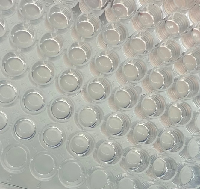
The simplest approach is 2D ECM coating. In this workflow, human ECM is applied to the surface of a culture plate before cell seeding. This allows researchers to add human matrix-derived cues into the culture environment. ECM coating can be useful for primary cells, attachment-sensitive cells, epithelial models, stromal cells and early assay optimization.
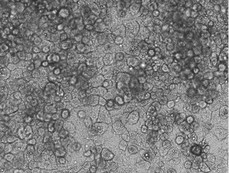
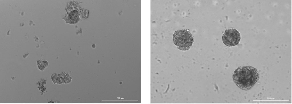
The second approach involves using human ECM in 3D spheroids or organoid-like models. Spheroids already improve model relevance by allowing cells to self-organize, form gradients and develop cell-cell interactions more similar to those in tissue. Adding ECM introduces another important layer: cell-matrix signaling. Creating 3D models based on ECM is less time-consuming compared to organoid systems, does not require activation of the Wnt-3a pathway and incorporates crosstalk between different cell types.
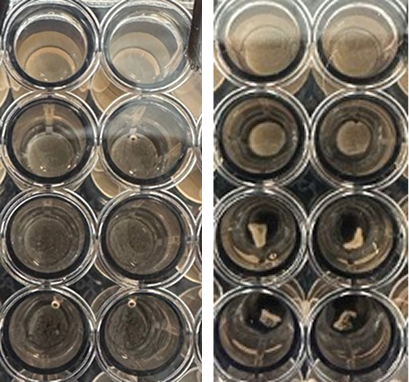
Human ECM can also support contracted disk models, where cells are mixed with ECM and allowed to self-organize into compact, contractile structures. These models are especially relevant for fibrosis research. Instead of measuring only cell viability, researchers can track outputs such as gel contraction, disk morphology, stiffness, matrix remodeling, cell viability and the expression of different markers.
What Human ECM solutions does Preci offer?
Preci offers a broad line of human-derived extracellular matrix products for building more physiologically relevant in vitro models. ECM from different human organs and tissue types is available, allowing researchers to select a matrix environment that better matches their biological question.
Each ECM batch is supported by quality control, including characterization of matrix composition and quantification of selected ECM proteins. Additional, project-specific quality control can also be requested.
For projects that require a more specific matrix environment, Preci can also develop custom ECM hydrogels. These can be produced from a selected organ, disease context, or donor group, depending on the research goal.
In addition to ECM products, Preci can provide donor-matched systems, where the matrix is combined with cells from the same donor. These may include blood cells, fibroblasts, epithelial cells, stromal cells or other cell types required for the model.
Preci also provides application protocols for different ECM workflows. When needed, these protocols can be optimized for a specific cell type, assay format, readout or drug development workflow.
Looking for a human matrix for your drug development workflow? Contact preci.bio to discuss Human ECM options, tissue-specific matrices, donor-matched cells and available protocols.
FAQ
Why is extracellular matrix relevant if the model already uses human cells?
Cells alone do not recreate a human tissue environment. Cell behavior is strongly affected by the surrounding matrix, including its composition, stiffness, architecture and bound signaling molecules. If the matrix is absent, oversimplified, or non-human, important tissue-specific signals may be missing.
How is human-derived ECM different from Matrigel?
Matrigel-like products are derived from EHS mouse sarcoma and are widely used in 3D culture. Human-derived ECM comes from human tissue, which makes it more relevant for modeling human tissue microenvironments, especially when tissue specificity or disease context matters.
Which disease areas benefit most from Human ECM-based models?
Human ECM is especially relevant for diseases where the microenvironment strongly affects cell behavior, such as cancer, fibrosis, chronic inflammation, wound healing and tissue degeneration.
Can Preci provide custom ECM for a specific project?
Yes. Preci can develop custom ECM hydrogels from selected organs, disease contexts or donor groups. Additional batch characterization and workflow optimization can also be requested depending on the project needs.
Written by Mariia Moshkivska