


Primary human preadipocytes are widely used to study adipogenic differentiation. However, adipose tissue remodeling extends beyond changes in lipid accumulation: under pro-fibrotic conditions, preadipocytes can also acquire myofibroblast-like features.
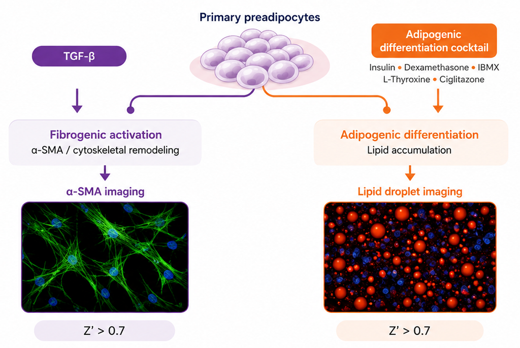
We therefore developed a preadipocyte fibrosis assay that combines two imaging-based phenotypic readouts in primary human cells: fibrogenic activation and adipogenic differentiation. This approach enables both responses to be evaluated across donor-derived cultures within the same experimental platform.
Assay setup
The assay was developed to screen primary human preadipocytes for their susceptibility to fibrogenic activation. It was evaluated using cells from two adipose tissue depots and from donors with and without diabetes:
| Donor | Depot | Donor status |
| EPI-02 | Epicardial | Non-diabetic |
| EPI-05 | Epicardial | Diabetic |
| PRE-02 | Visceral | Non-diabetic |
| PRE-05 | Visceral | Diabetic |
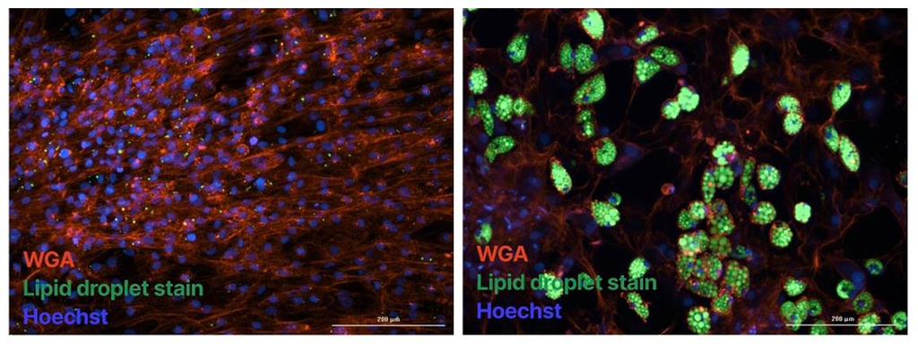
For the adipogenic arm, cells were differentiated using insulin, dexamethasone, IBMX, L-thyroxine, and ciglitazone. Lipid droplets, nuclei, and cell morphology were visualized by fluorescence imaging, and lipid-positive area was normalized to cell number.
For the fibrogenic arm, cells were exposed to TGF-β as pro-fibrotic stimuli. Fibrogenic activation was assessed by α-smooth muscle actin (α-SMA) immunofluorescence.
Establishing a robust fibrogenic readout
Secreted COL1A1 was initially evaluated as a readout of fibrogenic activation. However, the ELISA showed insufficient reproducibility across donor cultures because measured collagen levels were influenced by differences in cell number and donor-dependent proliferation.
We therefore moved to an imaging-based α-SMA readout, quantifying the percentage of α-SMA-positive cells rather than total secreted protein. This provided a more reproducible quantitative endpoint for comparing fibrogenic responses across donor cultures.
The α-SMA assay achieved a Z′ factor >0.7, indicating robust separation between assay controls and supporting its use for quantitative phenotypic analysis.
The assay reveals donor-specific responses
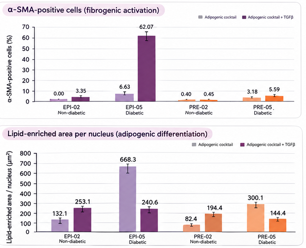
Seven days after pro-fibrotic stimulation, the assay resolved different α-SMA response patterns across the four donor-derived cultures.
| Donor | α-SMA response | Phenotype |
| PRE-02 | Minimal induction | Resistant |
| EPI-02 | Minimal induction | Resistant |
| PRE-05 | Measurable increase | Slightly inducible |
| EPI-05 | Strongest response; elevated baseline | Strongly inducible, high baseline |
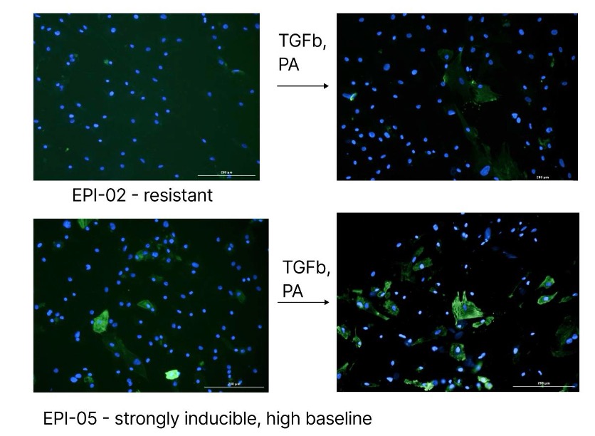
The assay captured distinct donor-specific fibrogenic responses, ranging from resistant to strongly inducible phenotypes. EPI-05 showed the strongest response to pro-fibrotic stimulation, together with elevated baseline α-SMA expression.
Adding adipogenic response as a second phenotype
Lipid accumulation was quantified in parallel as lipid-positive area normalized to cell number. Adipogenic capacity differed among donor-derived cultures, but TGF-β treatment did not consistently reduce lipid accumulation across all four models: a donor can show a strong fibrogenic response without a corresponding change in adipogenic differentiation. Measuring both endpoints within the same assay therefore captures phenotypic responses that neither readout would reveal alone.
What this assay enables
The established workflow provides a quantitative primary-cell platform for characterizing donor-dependent preadipocyte phenotypes and evaluating responses to pro-fibrotic stimulation.
- Validated preadipocytes with the largest assay window for fibrogenic activation
- Donor characterization and selection for downstream studies
- Quantitative assessment of TGF-β-induced fibrogenic activation
- Parallel measurement of fibrogenic and adipogenic phenotypes
- Compound testing in primary human preadipocytes
- Phenotypic studies in fibrosis and cardiometabolic research