

ECM proteome of cardiac tissue and cardiovascular disease
The human cardiac extracellular matrix (ECM) is a highly specialized, dynamic three-dimensional network of biomolecules that helps cardiac tissue maintain its structure and integrity and provides biochemical signaling and mechanotransduction to cardiac cells. ECM also coordinates many interrelated functions, including the storage and release of growth factors, delivery of intercellular communication, regulation of tissue repair and fibrosis formation, control of angiogenesis, modulation of inflammatory responses, and support for the propagation of electrical impulses of cardiomyocytes.
When the ECM’s structure, composition, or remodeling gets disrupted, it becomes a central driver of cardiovascular disease. Conditions like myocardial infarction, hypertensive stress, and dilated cardiomyopathy trigger ECM remodeling by activating fibroblasts and pushing them to transdifferentiate into myofibroblasts. This process is fueled by TGF-beta, mechanical stress, neurohumoral factors, inflammation, and shifts in matrix metalloproteinase activity. As a result, excessive deposition of fibrillar proteins accumulates in the myocardium, increasing stiffness and scarring (hallmarks of nearly all forms of heart failure).
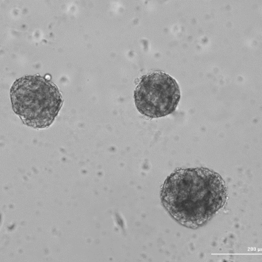
In addition to vivo studies, assays based on extracellular matrix are widely employed in laboratory research to investigate the mechanisms of cardiac remodeling and disease progression under controlled experimental conditions. The combination of heart cell populations with matching organ ECM hydrogels enables the establishment of physiologically relevant in vitro models that recapitulate key features of cardiovascular pathologies, including fibrosis, aberrant matrix remodeling, and altered cell–matrix interactions. To facilitate such applications, our company develops ECM-based hydrogel systems specifically designed for advanced in vitro modeling and extracellular matrix research.
This review summarizes common knowledge about the molecular composition of the cardiac extracellular matrix proteome and outlines the mechanisms that contribute to disease-associated extracellular matrix remodeling.
The Normal Cardiac Matrisome: Composition and Architecture
1. Collagens and Structural Proteins
If you think of the heart as a living machine, its extracellular matrix (ECM) is the structural framework holding everything together — and collagen is the steel of that framework. The two dominant fibrillar collagens in cardiac tissue are type I and type III, and their proportions matter enormously. Type I collagen accounts for 80–90% of total myocardial collagen and type III for approximately 11%, together providing both structural strength and a degree of elasticity to the heart wall. The cardiac collagen network is arranged hierarchically: large coiled perimysial fibers provide tensile stiffness, while smaller endomysial fibers surrounding individual cardiomyocytes prevent cell slippage and maintain ventricular geometry in the unloaded state.
Beyond these two dominant collagens, types IV, V, and VI are also present in the normal cardiac proteome. They serve basement membrane integrity, fibril nucleation, and flexible inter-cell linkage, respectively.
Elastin complements this framework by giving the ventricular wall its ability to recoil after each contraction, enabling efficient passive filling.
Fibronectin is a large dimeric glycoprotein that exists in at least 20 splice variants. It is critical for cell adhesion, migration, and matrix assembly, and plays an especially important role during disease. Laminins hook the cardiomyocyte cytoskeleton to the surrounding ECM via integrin receptors, forming the structural core of basement membranes.
2. Proteoglycans and Glycosaminoglycans
Collagen gives the cardiac ECM its mechanical backbone. Proteoglycans and glycosaminoglycans handle chemistry. They keep the matrix hydrated, they shape local inflammation, and they help carry signals between cells.
The heart relies on a handful of proteoglycans: versican, decorin, biglycan, lumican, aggrecan, perlecan, and the syndecans. Versican is the large one, rich in chondroitin sulfate. It affects how viscoelastic the tissue is and how easily inflammatory cells move through it. How fast it gets replaced comes down to the ADAMTS proteases. Decorin and biglycan sit in a different class, the small leucine-rich proteoglycans, or SLRPs. They bind straight onto collagen fibrils and set how thick each fibril grows and how far apart they sit. That, in turn, feeds back into how the whole network behaves under load. Decorin has one more trick. It grabs TGF-β, a cytokine that pushes fibrosis, and holds it tightly. With less TGF-β free to act, fibrotic signaling in the cardiac interstitium stays quieter.
Then come the glycosaminoglycans, or GAGs: hyaluronan, heparan sulfate, chondroitin sulfate, dermatan sulfate. All of them carry a negative charge. They draw water into the matrix and shape the local cytokine gradients. Hyaluronan reacts quickly to injury. After ischemia reperfusion is made within hours and collected in the infarcted left ventricle over the first day. Remove it, and things get worse: macrophage responses falter, and so does hemodynamic recovery. Its breakdown is where the problem starts. Hyaluronan is normally laid down as a high-molecular-weight polymer, and in that form it tends to be anti-inflammatory. Once it is cut into low-molecular-weight fragments, the effect flips: those fragments act as danger signals, prompting endothelial and immune cells to release cytokines and chemokines.
3. Matricellular Proteins
Some matrix proteins barely touch the structure. Their job is communication. They sit between a cell and its surroundings and relay information. This group, the matricellular proteins, includes osteopontin, periostin, tenascin-C, SPARC (secreted protein acidic and rich in cysteine), galectin-3, and thrombospondins.
Timing is what sets them apart. You will hardly find them in a healthy adult’s heart. Put that heart under stress, injury, or inflammation, though, and their levels climb fast. They do not reshape the matrix so much as steer the cells inside it. Working through integrins and growth factor receptors, they change how cardiomyocytes and interstitial cells read their surroundings and respond. Both resident myocardial cells and the ones that arrive during injury secrete them. Together, they help direct adhesion, migration, growth, and differentiation as tissue remodels.
4. Matrix Metalloproteinases and Their Inhibitors
The matrix is never finished. It gets torn down and rebuilt around the clock. The tearing down belongs to the matrix metalloproteinases, the MMPs: a family of more than 20 zinc-dependent endopeptidases, each one fussy about what it cuts. The heart carries several. Collagenases (MMP-1, -8, -13), gelatinases (MMP-2, -9), a stromelysin (MMP-3), and the membrane-bound types (MT1-MMP/MMP-14). Proteoglycans are a separate task, and that one falls mostly to the ADAMTS proteases, which cut versican and aggrecan.
The activity of MMPs is kept in check by tissue inhibitors of metalloproteinases (TIMPs 1-4). The MMP/TIMP balance is the central dial of ECM homeostasis, tipping it in either direction is pathological. Excessive ECM accumulation increases wall stiffness and impairs compliance, causing diastolic dysfunction. Whereas inadequate deposition and collagen assembly lead to progressive infarct wall thinning, ventricular dilation, aneurysm formation, and rupture.
5. Growth Factors and Other Matrisome Associated Proteins
One of the most underappreciated roles of the cardiac ECM is its function as a regulated reservoir of soluble signaling molecules. TGF-β isoforms, FGFs, VEGFs, and IGFs are all stored in latent, inactive form within the matrix: they are bound to proteoglycans such as decorin, biglycan, and perlecan; and released on demand in response to injury or proteolytic activation. Through this mechanism, GAGs and proteoglycans effectively sequester growth factors and cytokines in the interstitial space until physiological or pathological cues call for their release.
This “on-demand” release system means the ECM is not a passive structure. It is an active participant in fibroblast activation, angiogenesis, and cardiomyocyte survival signaling. In a healthy organism, the cardiac ECM does more than provide mechanical support. It actively transduces signals necessary for cell survival and function. Most of the cardiac pathologies involve expansion of the interstitial matrix and marked shifts in its composition, both of which disrupt systolic and diastolic function.
ECM Proteome in Specific Cardiovascular Pathologies
1. Myocardial Infarction and Post-Myocardial Infarction Remodeling
After myocardial infarction, the left ventricle undergoes a complex repair process whose success rate is tightly connected with ECM response. This process consists of three overlapping steps: an initial inflammatory burst, a rebuilding/fibrotic phase, and a final maturation stage.
In the first hours after the infarct, dying cells and activated MMPs tear apart the native collagen scaffold. A provisional matrix — built from fibrin and fibronectin — fills the void, acting as a temporary platform for immune cells and repair-competent fibroblasts to move in.
Fibronectin expression spikes dramatically after MI, and its different variants can lead to diverse outcomes. The fibronectin-EDA isoform acts as a kind of “danger signal” for immune cells, and mice lacking it showed significantly less left ventricular dilatation and better systolic function compared to wild-type animals, despite identical infarct sizes. Absence of FN-EDA enhanced survival and cardiac performance by modulating matrix turnover and inflammation through leukocytes and fibroblasts.
During the rebuilding phase, cardiac fibroblasts differentiate into myofibroblasts under TGF-β signaling and deposit large amounts of structural collagen to form a mechanically stable scar. Matricellular proteins — tenascin-C, periostin, SPARC, osteopontin, thrombospondins-1 and -4 — are all strongly induced and coordinate scar formation and collagen cross-linking.
Quantitative ECM proteomics using porcine ischemia-reperfusion models has uncovered several matrix proteins that accumulate specifically in the focal lesion. For the first time, proteins such as cartilage intermediate layer protein 1 (CILP1), asporin, adipocyte enhancer binding protein 1 (AEBP1), and collagen XIV were identified as contributors to cardiac scar remodeling after ischemia.
The ratio of matrix metalloproteinases (MMPs) to their tissue inhibitors (TIMPs) determines the trajectory of post-infarct remodeling. Excessive MMP activity risks ventricular rupture, whereas insufficient activity results in a rigid, non-compliant scar that promotes heart failure. While MMP-2, -9, and -14 drive this post-MI remodeling, a high MMP-9 to TIMP-1 ratio is most consistently linked to adverse left ventricular outcomes.
2. Heart Failure and Cardiac Fibrosis
It barely matters what triggers heart failure. The matrix changes the same way each time. Collagen and other ECM proteins pile up in the myocardium, the ventricle grows stiffer, and both filling and pumping get harder.
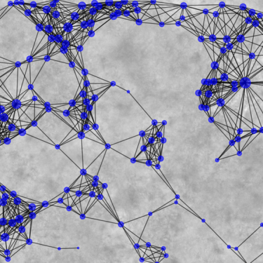
How far this can go came through clearly in a recent proteomic study of end-stage ischemic cardiomyopathy (ICM). Working on left ventricular tissue with high-resolution mass spectrometry, the team quantified 315 ECM proteins. LOXL1, FBLN1, and versican stood out as the most strongly upregulated. When the researchers traced which pathways were active, three lit up together: TGF-β signalling, integrin-mediated adhesion, and complement. That trio reads like a loop feeding itself, one that keeps the failing heart laying down still more matrix.
Step back to the wider literature and the count grows. ECM proteomics of ischemic heart failure has now logged more than 200 matrix-related proteins in human myocardium. Fibrillar collagen aside, the interstitial proteins that build up most are the SLRPs and versican (VCAN). Single-cell sequencing supports this, showing that versican and ADAMTS expression really are specific to the human heart. Part of the versican buildup traces to a single enzyme easing off: ADAMTS5 simply cleaves less of it. This is a clinically useful piece. Beta-blockers blunt that buildup, which ties an everyday neurohormonal therapy straight to what the matrix is made of.
Pressure-overload failure looks a little different. In hypertensive heart disease and aortic stenosis, proteins that had vanished from the healthy adult heart turn up again: fibronectin-EDA, tenascin-C, osteopontin, and the thrombospondins. None of them holds up the structure. They send signals instead, switching on hypertrophic gene programmes and fibroblast activity through integrins.
3. Atherosclerosis and the Vascular ECM
A healthy artery wall is built from fibrillar collagens, elastin, proteoglycans (versican, biglycan, decorin), and fibronectin. Atherosclerosis takes that apart and rebuilds it. The opening step is the decisive one. Apolipoprotein B-containing lipoproteins get caught in the subendothelial matrix, snagged by biglycan and versican, and that is what seeds the plaque.
Matricellular proteins you would struggle to find in a healthy vessel show up in force inside a lesion. Osteopontin, tenascin-C, SPARC, thrombospondin-1: each one nudges smooth muscle cells to migrate and pulls in inflammatory cells. At the same time, the proteases lose their balance. MMP-1, -3, -8, and -9 climb while the TIMPs drop, and the fibrous cap thins until the plaque sits one bad moment from rupture.
Valves are made differently from the myocardium, with an ECM arranged in layers. Proteomic studies in patients undergoing valve surgery have turned up disease-specific ECM signatures. These may help with a puzzle that has lingered in the clinic for years: why the left ventricle recovers more dependably after surgery for aortic stenosis than for aortic regurgitation. Nobody has fully explained it yet.
4. Valvular Heart Disease
Calcific aortic valve disease brings its own mix of changes. Collagen turns fibrotic, elastin breaks down, and mineral settles where it should not. The main drivers of that calcification are SPARC, osteopontin, osteocalcin, and the bone morphogenetic proteins.
Conclusion
For a long time, the cardiac extracellular matrix was treated as little more than scaffolding. That view has changed. Thanks largely to high-resolution proteomics, it now reads as an active part of how the heart works, and one you can actually measure. Look across myocardial infarction, heart failure, atherosclerosis, and valvular disease, and the same lesson keeps surfacing. Disease rarely hangs on one protein. It comes from many shifts at once: what the matrix contains, and how quickly it is replaced. A few of those shifts already matter at the bedside. The MMP-to-TIMP balance steers how a heart remodels after infarction, and beta-blockers hold versican back in failing hearts. Findings like these make the ECM a credible place to hunt for biomarkers and drug targets. The rest should follow from pairing two things: proteomic readouts from human tissue, and defined ECM-based models that let researchers test, under controlled conditions, which matrix changes truly drive disease, and how they might be reversed.
Written by Yelyzaveta Kononenko